One of the great challenges of the last twenty years for the study and conservation of cultural heritage is the development of non-invasive investigation techniques, i.e. techniques and devices capable of revealing the state of conservation of an artwork without damaging it, along with the technique and materials used by the artist. Works of art are both unique and extremely delicate objects, handed down through the history of art and humanity. This is where photonics, a branch of optics and physics, comes to our aid. In recent decades, optical and spectroscopic devices have been developed, getting more precise, portable, and smaller, which can provide a host of information about the materials that go into the composition of an artwork.
To help us understand such complex techniques, we asked Professor Daniela Comelli, a lecturer at the Polytechnic and the head of ArtIS (Imaging and Spectroscopy for Art) research group of the Physics Department.
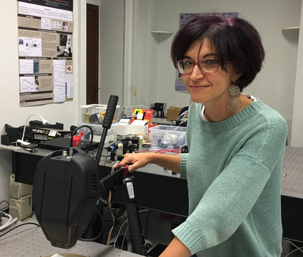
The laboratory focuses its research on the development of spectroscopy and imaging methods for the non-invasive study of artworks. Time-resolved fluorescence imaging, hyperspectral imaging and Raman spectroscopy are some of the techniques used. Never fear, we shall explain all these terms shortly.
The research group has done work on extraordinary artefacts, including one of the daggers found in King Tutankhamun’s sarcophagus, an illuminated manuscript from the Trivulziana Library collection in Milan, two of Michelangelo’s most famous marble sculptures—David and Rondanini Pietà—, and modern art paintings such as Van Gogh’s “Les Bretonnes et le pardon de Pont Aven” and Pablo Picasso’s “Femme (Époque des ‘Demoiselles d’Avignon’)”.
How did your passion for physics applied to the world of art arise?
I started my thesis in a very different field of application, biology and medicine, where I used the same imaging spectroscopy techniques that I use today to study works of art. At that time, going on twenty years ago, we were analysing lab mice on which we inoculated tumours for the purpose of studying anti-tumour therapies. In the last year of my PhD, I moved on to the art world, and I liked it so much that I decided to stay.
What difficulties do you encounter when dealing with artworks?
Artworks involve a complex use of materials. I have come to understand that analysing a work of art implies dealing with various materials, which often overlap and sometimes have become degraded from the time when the artist used them. Because of this complexity, it is essential that we use a “multi-analytical” approach, that is, different but complementary investigation techniques, so we may understand what lies behind a work of art. Sampling may be required, and some of the techniques may be invasive or destructive.
How can you achieve less invasive techniques?
There is now great interest in the development of non-invasive techniques, which will not cause any damage to the work and can be achieved using portable tools. An artwork rarely every comes to us, it is generally we who go to where the artwork is to perform our analyses. Art conservators and curators always ask us many questions, such as what technique and materials the artist used, and whether there are ongoing degradation processes.
Let’s start on our journey with the dagger in Tutankhamun’s sarcophagus…
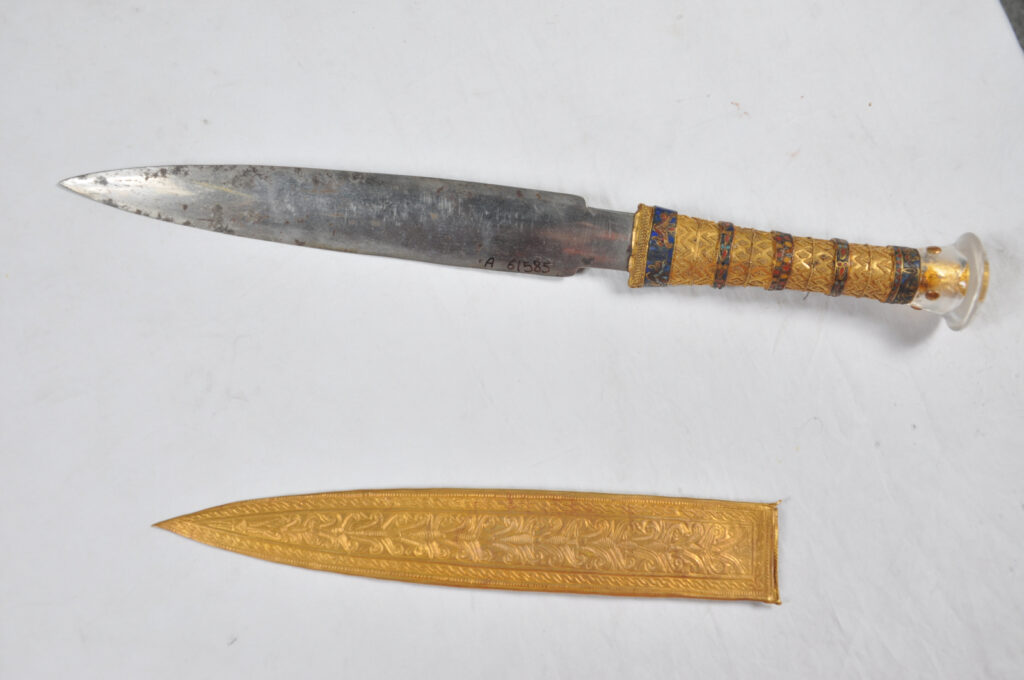
We shall start our exploration in ancient Egypt. We were given the opportunity to study one of the two daggers found in the sarcophagus of King Tutankhamun, who lived between 1341 and 1323 BCE. The research was long and complex, but it is certainly one of the most fascinating Egyptology studies we have ever done.
We are in the Amarna Period, fourteenth century BCE. Besides the ancient Egyptians, other important civilizations of that time were the Hittite empire, the Mitanni, and the Assyrians. Tutankhamun’s grandfather was the pharaoh Amenhotep III, who had left behind a very prosperous and peaceful Egypt, thanks to a treaty with the neighbouring Mitanni. His successor was Amenhotep IV, who had brought about big changes, especially in the sphere of religion, replacing the god Amun and all the other gods with a single deity named Aten, who was represented by the Sun, a sort of monotheism. Amenhotep IV and his wife were worshipped as direct emanations of Aten, such that the pharaoh changed his name to Akhenaten, meaning “the one who acts on behalf of Aten”, and moved Egypt’s religious capital from Thebes to the new city of Akhetaton, the present-day Amarna.
His son Tutankhaton took the throne while still a child, but soon changed his name to Tutankhamun, “living image of Amun”, as he restored the polytheistic religion of Amun and the other gods, even going as far as demolishing the temples dedicated to Aten. He was certainly a much-loved pharaoh, but he died very young, at only 19 years of age.
The discovery of his tomb was quite an event at the time …
Archaeologist Howard Carter led the research, which went on from 1915 to 1922. It was a grandiose tomb, which contained more gold than all the Egyptian banks at that time.
It took three years to open the sarcophagus. Carter found some precious objects wrapped in the bandages of the mummy, gifts for the pharaoh venturing into the afterlife, among which were two daggers, one with a gold blade and the other apparently with an iron blade. The latter immediately intrigued both Carter and the other Egyptologists of that time, for there was hardly any rust on it even though three thousand years had passed.
What was intriguing about that dagger?
Iron, which the blade appeared to be made of, was not yet produced as a material at the time Tutankhamun was alive, which was still during the Bronze Age. The Iron Age began after 1200 BCE in Asia Minor, and a little later in Egypt. Tutankhamun’s dagger is not the only iron object dating back to a historical period prior to the Iron Age; more have been found, including from other civilizations. In all these cases, the question is whether the iron’s origin is meteoritic or terrestrial, obtained by chance during the smelting of bronze. What is certainly true is that, at the time of Tutankhamun, objects made from iron were so rare that they were considered extremely valuable, even more than objects made from gold.
The question was therefore to investigate the origin of the iron: was it meteoritic or quarry earth iron?
Yes, and it is a long story. As early as 1973, the researcher Bjorkman hinted at a possible meteoritic origin for the iron of the dagger blade but without backing his claim with scientific evidence. Then, in 1994, an investigation with X-ray fluorescence spectroscopy (XRF spectroscopy) indicated that the metal was not of meteoritic origin. The plot thickened in 2010, when researchers from the Italian city of Pisa discovered that the Egyptian Kamil crater had been created by the impact of a ferrous meteorite, which led to the hypothesis that the ancient dagger could have been made with fragments of that iron meteorite.
What scientific methodologies can help us unravel this mystery?
Meteoritic iron can be distinguished from terrestrial iron primarily by its microstructure, but a micro-sampling analysis cannot be performed on the precious dagger that belonged to King Tutankhamun.
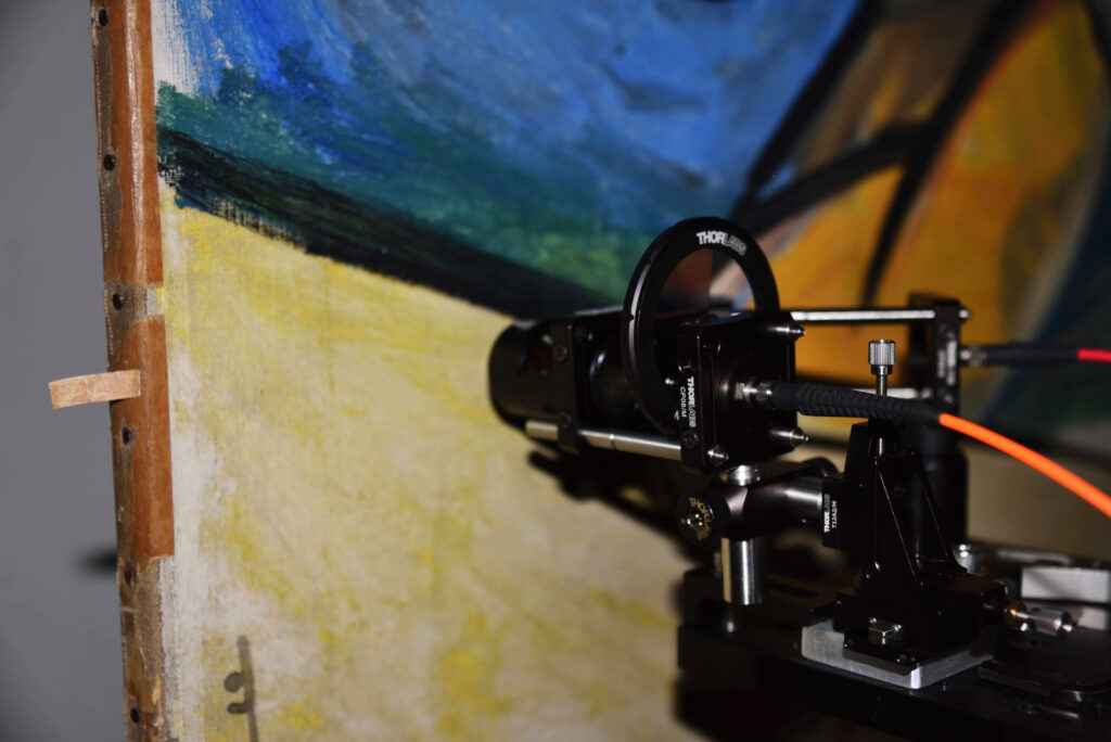
However, with the X fluorescence spectroscopy technique, we can study the chemical composition of iron and establish the presence of elements typical only of meteoritic iron, with the great advantage of using portable instruments.
This technique consists in irradiating the material with high-energy incident radiation, typically X-rays, which causes an atom to expel electrons from the innermost orbitals. As electrons are ejected, they leave a gap, which causes a state of imbalance whereby the electrons of the outermost orbitals are forced to make an energetic leap to fill the gap, thus releasing a unique type of energy that can be detected to identify the material. By recording the energies emitted, we can obtain an X fluorescence spectrum, showing very precise peaks, each of them associated with a chemical element. Thus, this technique allows us to determine the chemical elements of a material.
In December 2004, we used this investigation technique on the dagger blade at the Egyptian Museum in Cairo. We were thus able to determine that the blade was made from iron as well as a high percentage of nickel (approximately 10%). This high percentage of nickel, together with the presence of trace amounts of cobalt, clearly indicate that we are dealing with meteoritic iron. At this point, we can say that Tutankhamun’s dagger was indeed made with fragments of a ferrous meteorite, although probably not from the Kamil crater.
Another interesting indication is the existence of a hieroglyph dating back to around 1300 BCE, “bia-n-pet”, which means “iron from the sky”, suggesting that the ancient Egyptians knew about these metal fragments from heaven, to be considered as a precious gift worthy of belonging to a much-loved pharaoh like Tutankhamun.
A question remains as to who made Tutankhamun’s dagger
Egyptian diplomatic documents such as the letters from Amarna tell of how the king of Mitanni had sent precious iron objects to Tutankhamun’s grandfather Amenhotep III. The letters list iron blade daggers and an iron and gold bracelet, therefore, we think that the dagger found in Tutankhamun’s sarcophagus had been a gift to his grandfather from the nearby Mitanni civilization. A recent study seems to confirm this hypothesis; the analysis of the dagger’s hilt in addition to the blade indicate that the materials were combined using a technology that was not typical of the Egyptian civilization in those times.
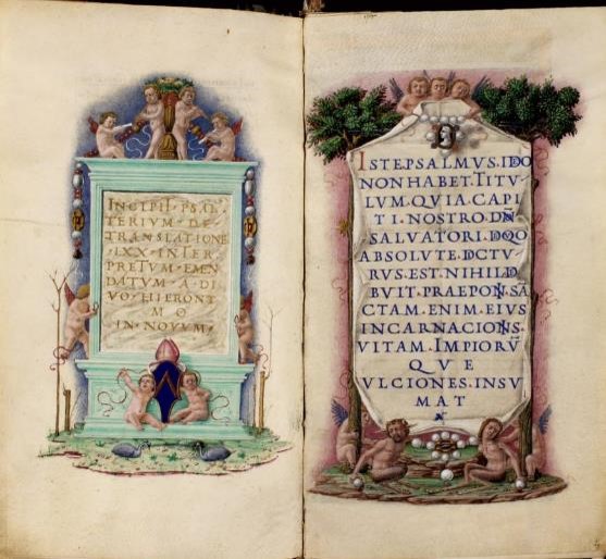
Let’s go back to the Trivulziana Library in Milan…
At the curators’ request, we analysed a small coat-of-arms from a fifteenth-century manuscript called Psalter. Finding a lesser quality design in such a well-crafted manuscript seemed odd to the curators, as the function of a coat-of-arms is to indicate the book’s owner, so they were wondering if it might have been repainted.
What techniques did you use in this case?
We used three non-invasive techniques: infrared reflectography, X-ray fluorescence spectroscopy, and Raman spectroscopy.
Infrared reflectography involves illuminating a surface with infrared light, such as from a common halogen bulb, and inspecting it with an infrared radiation camera. Infrared radiation can penetrate deeper layers of paint than visible radiation, so it is generally used to bring to light the preparatory drawing of a painting, pentimenti during the creation process, anything that might be found under the overlying pictorial layer.
Infrared reflectography revealed an underlying design in the likeness of a lion. However, colours are a very important aspect in heraldry, which means that a coat-of-arms depicting a lion may belong to different families depending on the colours used.
To determine what pigments were used in the underlying layer, we examined it with X fluorescence to map its chemical elements and Raman spectroscopy to identify its molecular compounds.
What were you able to find out?
The combination of the two spectroscopic techniques show that the overlying design is not original; it was made with ultramarine blue and red lead pigments. As for the underlying symbol, it features a bordure made with cinnabar, a red, mercury-based pigment, and a white lion made with white lead pigment on a blue background made with azurite.
By identifying the colours, we managed to recognize Bishop Bernardo de’ Rossi’s coat-of-arms, as shown on the cover of his portrait. Therefore, Bishop de’ Rossi was the original owner of the manuscript.
Tell us about your study on the stained-glass windows of Santo Spirito Church in Milan
In this case, we used a very interesting technique, called hyperspectral imaging. Developed and optimised over the last 20 years, this technique is currently used in various fields of application, like biology, medicine, industrial food contamination, and even remote vegetation monitoring from satellites and airplanes. The technique has been used in recent years for cultural heritage diagnostics applications.
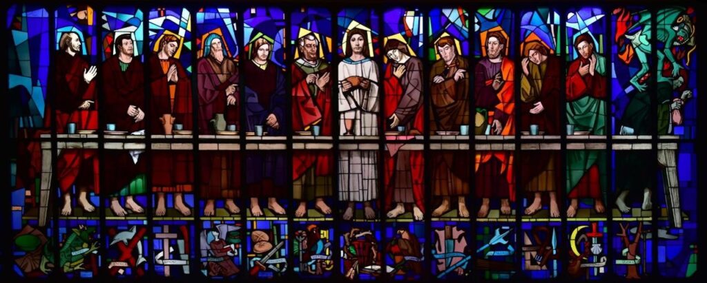
To start with, a black and white image is a single band image, whereas a colour image from a camera is made up of three bands called RGB colours (red, green, and blue). Hyperspectral imaging multiplies the light spectrum from three to about one hundred bands, combining the simplicity and immediacy of an imaging technique with the potential of a spectroscopy technique.
We did not use any lamps on the beautiful stained-glass windows of Santo Spirito Church, we just took advantage of a wonderful sunny day. Using a hyperspectral camera, we observed the different light spectrum responses transmitted by the colours from the glass, just as we do with our eyes when we recognise colours.
However, the hyperspectral camera showed that apparently identical pieces, blue, for example, had different transmission spectra and so were different from each other. You cannot see that with the naked eye because the phenomenon occurs in the near infrared (NIR). Our conclusion is that some of the glass pieces had probably been replaced at some point.
Let’s turn to the theme of modern painting…
The period from late 1800 to early 1900 is very interesting from the point of view of new materials available to painters.
In 1841, an American portrait painter by the name of John Rand came up with the idea of soft metal tubes to hold colours, the same in use today, replacing the pig bladder packets where colours were being kept until then, which were unable to prevent the colours from drying out quickly. The Impressionists found this novelty all-important, as they loved to paint outdoors, and indeed all the painters who followed them. The French impressionist Renoir observed that “without the paint tube there would have been no Cézanne, Monet, Sisley, or Pissarro, nothing of what journalists would call Impressionism”. The same is probably true for Renoir himself.
Another innovation came during that period in the form of modern pigments. These were created by means of chemical synthesis thanks to the chemical industry brought about by the Second Industrial Revolution. Examples of these modern pigments are zinc white, cadmium yellow and red, and chromium yellow. The chromatic brilliance and covering power of these colours were much higher compared to traditional pigments, which is the reason why the painters of that time used them intensely. It turned out only later that some formulations of these pigments were not very stable, for the synthesis methods used to make them had not yet been perfected.
As a result, some degradation phenomena are a typical occurrence in modern painting, such as protrusions, cracks, exudations, and fading.

We were given the opportunity to study these types of degradations on “Femme”, a painting by Picasso currently kept at the Bayeler Foundation in Basel, Switzerland. The painting dates from 1908 and is probably a study for one of his most famous works, “Les Demoiselles d’Avignon”. We compared two photographs of “Femme”, one taken in 2014 and the other in 1965, showing that the yellow coats kept their colour in the central part of the painting, but had clearly turned to a brown tone and were more faded on the sides.
We ran a series of spectroscopic and imaging analyses right on the painting as well as two microscopic fragments of paint taken from it. Our analyses showed that the two yellow coats were made with the same cadmium yellow pigment, but probably obtained from two tubes of different quality. As a result, the two cadmium yellows from different tubes changed in different ways, although they had been exposed to the same environmental conditions during the life of the painting; one has remained stable over time, the other is clearly degraded.
How did you determine the difference between the two paints?
We used the technique of investigating the optical fluorescence emitted, by which we illuminate a painting with ultraviolet light and then we photograph the radiation emitted back by the painting. Degraded yellow has an almost orange emission, while faded yellow has a much weaker emission and darker colour. This tells us that there are different materials, although both are cadmium yellow.
Indeed, other modern paintings made with cadmium yellow have shown the formation of a similar orange fluorescence when degradation is present. Therefore, we asked ourselves what was causing the orange fluorescence, whether it might be a useful indicator of cadmium yellow degradation and, last but not least, why some cadmium yellow formulations are more stable than others.
What is the origin of this orange emission?
We measured the optical emission again, to see how long it would last, knowing that fluorescence lasts a shorter time than phosphorescence. A detector showed us that it lasted for a few microseconds, which looked more like phosphorescence, much more intense in the degraded yellow area. Hence, we realized that defects in the crystalline structure of the pigment induced an emission from “trap states”.
Please tell us more…
There are various shades of Cadmium yellow, which is based on the semiconductor cadmium sulphide; lighter shades are obtained by adding zinc inside the crystalline structure, while darker shades are obtained by adding selenium. Interestingly, the paintings that show this kind of degradation all seem to come from the same historical period when the synthesis process of this colour was in its early stages and likely not yet perfected.
Cadmium yellow is a semiconductor in which sulphur and cadmium atoms alternate in a precise crystalline structure. However, when a crystalline material is synthesised, it is natural for defects to form inside the structure, caused by the synthesis process itself as well as interactions with other materials.
These defects create energy levels called trap states, which can give rise to an optical emission just like the one from Picasso’s painting.
Our conclusion is that the degraded coating is indeed made from cadmium sulphide, but with a much higher presence of crystalline defects than in the non-degraded coating, which defects produce an intense “phosphorescence” emission.
We looked at the literature to find an explanation for the orange emission from the degraded yellow; apparently, the smaller the cadmium sulphide crystals, the more the emission energy moves downwards from infrared to red-orange. We think that the cadmium yellow in question contains nanocrystalline particles of cadmium sulphide, which are much more reactive nanoparticles characterized by high-density surface defects and more easily subject to degradation. This hypothesis is also supported by the study of degraded cadmium yellow in Munch’s “The Scream”.
The research process must have been quite complex…
This last research is probably the most complex of those I reported. Despite its complexity, it brings us closer to the fascinating world of crystalline solids and defects in crystalline structures, a field of study that reminds me of a beautiful phrase by Colin Humphreys:
Crystals are like people: it is the defects in them which tend to make them interesting!
Crystals are like people: it is the defects in them which tend to make them interesting!